Rapid Identification of Stacking Orientation in Isotopically Labeled Chemical-vapor Grown Bilayer Graphene by Raman Spectroscopy
- Category: Materials, Nanotechnology
- Tags: jing kong, wenjing fang
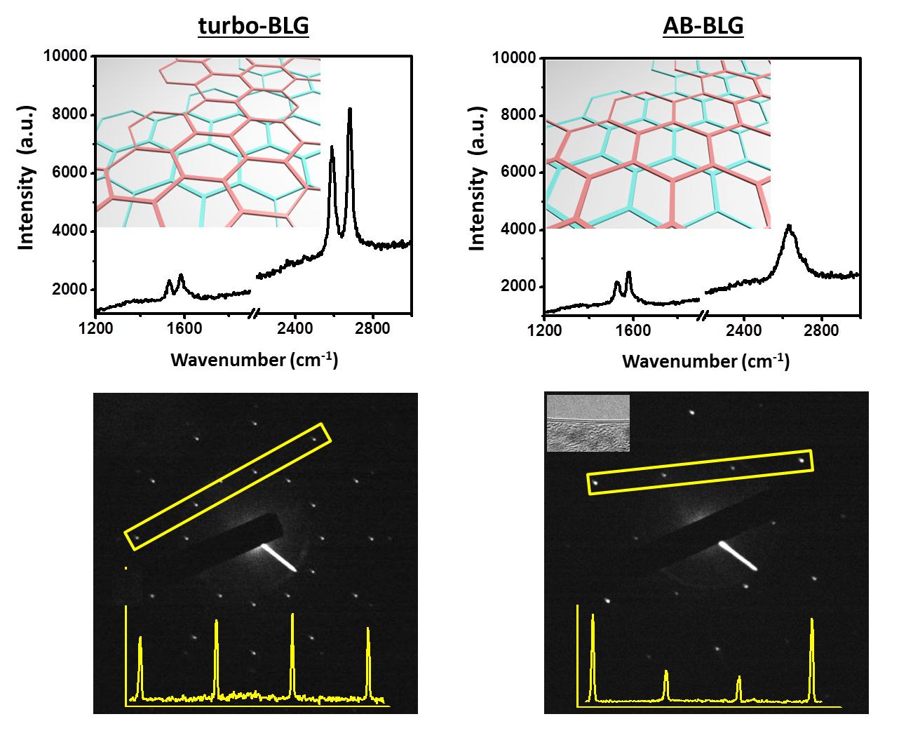
Figure 1: Raman spectroscopy and TEM characterization of bilayer graphene. (a) Two representative Raman spectra taken from suspended bilayer graphene on a SiN TEM grid. The insets show the stacking orientation. (b) The SAED patterns for (a) and the corresponding intensities profile along the yellow lines. Inset in upper-left corner of (b) is TEM image, which indicates the number of layers.
The growth of large-area bilayer graphene has been of technological importance for graphene electronics. The successful application of graphene bilayers critically relies on the precise control of the stacking orientation, which determines both electronic and vibrational properties of the bilayer system. Toward this goal, an effective characterization method is critically needed to allow researchers to easily distinguish the bilayer stacking orientation (i.e., AB stacked or turbostratic). In this work, we developed such a method to provide facile identification of the stacking orientation by isotope labeling. Raman spectroscopy of these isotopically-labeled bilayer samples shows a clear signature associated with AB stacking between layers, enabling rapid differentiation between turbostratic and AB-stacked bilayer regions. Using this method, we were able to characterize the stacking orientation in bilayer graphene grown through low-pressure chemical vapor deposition (LPCVD) with enclosed Cu foils, achieving almost 70% AB-stacked bilayer graphene. Furthermore, by combining surface sensitive fluorination with such hybrid 12C/13C bilayer samples, we are able to identify that the second layer grows underneath the first-grown layer, which is similar to a recently reported observation.