Flexible Probes for in vivo Recording and Stimulation of Neural Tissue
Probing and stimulating neural tissue is important to understand fundamentals of information processing and pathologies of the nervous system such as Parkinson’s disease, epilepsy, or spinal cord injury. Therefore it is desirable to collect data from multiple recording sites with high resolution and specifically manipulate neuronal activity in the regions of interest. Moreover, unspecific interaction with the probed tissue should be reduced to a minimum.
Current technologies to achieve this collectionrecord and stimulate the nervous system are usually based on materials such as silicon, silica, or metals, which suffer from fundamental physical limitations: low signal-to-noise ratio, low resolution, and deterioration of the quality of recordings[1].
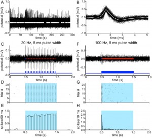
We present a novel method to fabricate devices for high-resolution recording, optogenetic stimulation, and pharmacological interrogation of neural tissue. In contrast to lithographically fabricated devices based on silicon or metal wires, our method results in highly flexible and more biocompatible devices, addressing the need for long-term recordings with limited interaction of implanted probes with the tissue. Employing a thermal drawing process inspired by optical fiber production, we create a scalable tool combining recording and intervention in neurophysiology. Our initial experiments demonstrate single-neuron recording and optogenetic stimulation of activity in the medial prefrontal cortex in vivo (Figure 1).

Figure 1: Neural recordings obtained from the medial prefrontal. (A) Activity in absence of stimulation. (B) Spike shapes isolated from data in (A). (C) Response to 20-Hz optical stimulation. (D) The multi-unit spikes in response to optical pulses across 20 trials identical to the one shown in (C). (E) Firing rate averaged across 20 trials shown in (D). (F) Response to 100-Hz optical stimulation. (G) The multi-unit spikes in response to optical pulses across 20 trials identical to one shown in (F). (H) Firing rate averaged across 20 trials shown in (G).
- M. P. Ward, P. Rajdev, and C. Ellison, “Toward a Comparison of Microelectrodes for Acute and Chronic Recordings,” Brain Research, Vol. 1282, pp. 183-200, 2009. [↩]