Nanostructured Photocathodes for Compact Coherent X-ray Sources
- Category: Electronic Devices, Nanotechnology, Optics & Photonics
- Tags: karl berggren, yujia yang
Nanostructured photocathode arrays can be used as the electron sources for the compact coherent X-ray source. Femtosecond laser pulses were used to actuate the photocathodes, resulting in ultra-short, high brightness and low emittance electron bunches. Subsequent electron-optic manipulations of these spatially periodic electron beamlets ready them for coherent X-ray generation via inverse Compton scattering[1].
We are interested in metallic nanoparticles as electron emitters because their plasmonic resonance can enhance the optical field by a factor of several tens and improve the electron emission yield of photocathodes. Moreover, the electron-optics of the compact coherent X-ray sources requires the pitch of electron emitters to be the X-ray wavelength multiplied by a magnification factor and the size of electron emitters to be less than a quarter of the pitch. For 10 keV X-rays and 1000 times magnification, the pitch is about 100 nm, and the size should be less than 25 nm. Therefore, we used high-resolution electron-beam lithography (EBL) and a metal lift-off process to fabricate the high-density, plasmonics-enhanced, nanoscale photocathode arrays. Au nanorod arrays partially embedded in a 10-nm SiO2 film were fabricated using a positive-tone EBL lift-off process, which employs ZEP-520A resist and a low temperature (0 °C) development procedure. The thin SiO2 film was used to block the photo-induced electrons emitted from the substrate.
Figure 1 displays the scanning electron microscopic (SEM) image of the Au nanorod photocathodes. The diameter and height of individual Au nanorod are 10 nm and 30 nm, respectively. This specific geometry tunes the plasmonic resonance band to around 800-nm free space wavelength to match the driving Ti:sapphire laser (800-nm wavelength) and maximize the optical field enhancement. Figure 2 displays the electron-emission yield from an array of Au nanorods. Electron emission current was measured as a function of incident laser power.
-
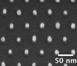
- Figure 1: SEM of Au nanorod array. The nanorod geometry was tuned to match a longitudinal surface plasmon mode of an Au nanorod, which was excited by 800-nm incident laser beam.
-
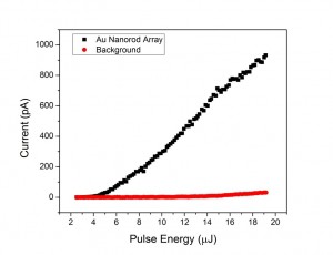
- Figure 2: Plot of emission current versus laser pulse energy for a 600-μm × 50 μm array of Au nanorods at 100-nm pitch, illuminated by 35 fs pulses of 800-nm light at a repetition rate of 3 kHz. Background emission is well suppressed by the thin SiO2 layer and photocurrent reaches 1 nA with 20-μJ incident laser pulse.
- W. S. Graves, F. X. Kärtner, D. E. Moncton, and P. Piot, “Intense superradiant x rays from a compact source using a nanocathode array and emittance exchange,” Physical Review Letters, 108, 263904, 2012. [↩]