Concomitant Co-crystallization on Engineered Surfaces
- Category: Materials, Nanotechnology
- Tags: allan myerson, xiaochuan yang
A co-crystal is a crystalline material made up of two or more components. In pharmaceutical systems, co-crystallization is frequently used to enhance the physical properties (e.g., solubility or melting point) of a drug substance via non-covalent interactions with a co-former. Numerous high-throughput screening methods have been proposed[1]. However, none of these studies explored the polymorphism of co-crystal. In our previous work[2], patterned self-assembled monolayers (SAMs) were shown to be an attractive tool for polymorph screening. To expand the application to co-crystallization, the patterned SAMs substrates were used for the concomitant co-crystallization of caffeine-oxalic acid.
The bi-functional patterned SAM was prepared at MTL (MIT) by photolithography using silicon wafer as the base substrate coated with 5-nm titanium and 50-nm gold layers. The patterned gold substrates have an island size of 500 µm, with the hydrophilic gold island functionalized with the –COOH group and hydrophobic OTS on the silicon substrate. Co-crystallization experiments were conducted by forming an array of droplets of saturated solution of caffeine and oxalic acid (2:1 molar ratio) in chloroform: methanol (7:2 v/v) onto the SAMs. Then, the solvents were allowed to evaporate slowly in a desiccator. Once the co-crystal was formed, the crystals were characterized by Raman spectroscopy and X-ray diffraction. Compared to the Raman spectrum of the co-crystal (reference) reported earlier[3], polymorphs with distinct new Raman peaks at 1076 and 1588 cm-1 were discovered. With six repeated experiments, more than 50% of the islands were found to be the reference co-crystal. In other (non-reference) islands, most of the co-crystals have peaks at 1076 and 1588 cm-1, with some of them having a lone peak at either 1076 or 1588 cm-1. In addition, the XRD diffraction pattern showed additional peaks at 2θ of 30o and 32.3o, further confirming the presence of a new co-crystal form.
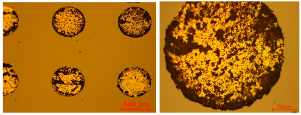
Figure 1. Co-crystals growing on SAMs at increasing magnification.
- M. A. Elbagerma, H. G. M. Edwards, T. Munshi, M. D. Hargreaves, P. Matousek, and I. J. Scowen, Crystal Growth & Design, 2010, 10, 2360-2371 (DOI:10.1021/cg100156a). [↩]
- A. Singh, I. S. Lee, and A. S. Myerson, Crystal Growth & Design, 2009, 9, 1182-1185 (DOI:10.1021/cg801055x). [↩]
- A. V. Trask, W. D. S. Motherwell, and W. Jones, Crystal Growth & Design, 2005, 5, 1013-1021 (DOI:10.1021/cg0496540). [↩]