Computational Electromagnetics Tools for High-field Magnetic Resonance Imaging
- Category: Medical Electronics
- Tags: jorge fernandez villena, luca daniel
Last-generation magnetic resonance imaging (MRI) scan technology allows combining higher fields with parallel transmit-coil arrays to improve the signal-to-noise ratio (SNR) while minimizing human-body heating by electric fields. However, these higher fields imply higher-frequency RF pulses, with wavelength comparable to the human body’s dimensions, which, besides degrading SNR factors, may create local hotspots in the body. Coil and pulse designs must comply with ruling safety regulations in terms of local and global specific absorption ratio (SAR) limits while providing better possible accuracy. Addressing such problems requires more sophisticated computational electromagnetics (CEM) tools for detailed analysis, a scenario that challenges state-of-the-art EM analysis tools. A lack of suitable analysis tools prevents exploitation of the full potential of the available technology. In close collaboration with the RLE MRI group and the Harvard Massachusetts General Hospital MRI group, the computational prototyping group is developing CEM techniques to address these new needs of the MRI community, working with specific targets.
The first target is developing efficient EM tools for complete analysis of highly complex highly inhomogeneous human body models. We have implemented a solver based on a new formulation combined with fast volume integral equation approaches to solve high-resolution models in less than a minute. The second target is combining volume and surface integral equation methods to efficiently model the complete coil array configuration and human body. The resulting models allow us to solve the complete coil and human body EM problem, including obtaining the coils’ port impedance needed for circuitry tuning and the electric and magnetic fields distribution in the body, thus efficiently predicting imaging quality and localizing potential bodily hotspots due to large electric field regions. To further speed up the analysis, this hybrid approach will combine pre-computed Green’s functions for a realistic human body model with a method of moments to rapidly assess different coil configurations for a typical body and thus allow for efficiently optimizing the geometrical configuration of the transmit coils. To aid this assessment, we plan to leverage our work on parameterized model-order reduction, automatically generating models depending on relevant parametric quantities. We are also working on applying the recently developed fast methods for computing the approximate solutions to the electromagnetic fields inside the body, assuming a simplified 3-tissue model, which can be obtained for each patient by a quick MRI scan. Such an approach will open the possibility of performing patient-specific on-line EM analysis to optimize the coil and pulses applied to obtain the best imaging for each patient’s anatomy.
-
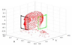
- Figure 1: Interpolation points used as pre-computed Green’s function for a given human body. This model can be used to speed-up the analysis of multiple coil array configurations.
-

- Figure 2: Sagittal view of 2.5-mm resolution Duke model. Top figures are real and imaginary part or relative permittivity. Bottom figures are the currents and fields generated when excited by a plane wave at 7T.