Point Spread Function for PMMA as a Negative Resist

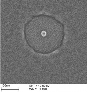
Figure 1: SEM image of a beam spot at 10 keV and a dose of 760 fC. The hole's diameter from the positive-tone response of the PMMA is 215 nm, and the diameter of the center pillar, produced by the negative tone PMMA, is 27 nm.
Poly(methyl methacrylate) (PMMA) is an important resist for electron-beam lithography. For low doses, it is a positive resist; for high doses, a negative resist. Knowledge of the point-spread function (PSF) is critical for proximity effect corrections (PEC) using electron-beam lithography with PMMA as a negative-tone resist [1]. An example of the positive- and negative-tone PMMA is shown in Figure 1.
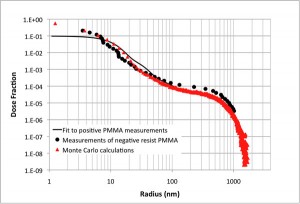
Sequences of single-point exposures are made in 40-nm PMMA at doses high enough to produce the negative resist behavior for electron energies of 2-30 keV. Development at 30°C in a mixture (2:1) of isopropyl alcohol and methyl isobutyl ketone yields center posts increasing in diameter with increasing dose. The PSF is determined from these measurements. The results for 10-keV electrons are shown in Figure 2.

Figure 2: Measurements and Monte Carlo calculations of the point spread functions for 10 keV electrons on 40 nm-thick PMMA. The PSFs for the positive- and negative-tone PMMA are scaled to overlay the normalized Monte Carlo PSF values. The fit to the positive-resist data is used for clarity.
The PSF function is usually modeled as the sum of two or more Gaussian functions. One Gaussian represents the effects of forward-scattered electrons and another represents the back-scattered electrons. Four Gaussians are used to model the data in Figure 2. The fit for the forward scattering is not well determined from the data because a 4.5-nm radius is the smallest feature size reliably measured.
A Monte Carlo simulation, including the effects of secondary electrons, is in good agreement with the measured PSF of Figure 2. It is used to improve the fit for forward scattering. The good agreement between the Monte Carlo simulation and the positive and negative resist PSFs in Figure 2 suggests that there is no significant difference in the physical interactions of the electrons with the PMMA resist to produce a positive- or a negative-tone effect other than total dose. The differences visible in the measured PSFs may be primarily due to a change in the density of the resist with dose, an effect not included in the Monte Carlo model.
References
- H. Duan, J. Zhao, Y. Zhang, E. Xie, and L. Han, “Preparing patterned carbonaceous nanostructures directly by overexposure of PMMA using electron-beam lithography,” Nanotechnology, vol. 20, 135306 (8pp), 2009. [↩]