Light-proof Electrodes for In-situ Monitoring of Neural Function

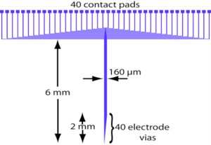
Figure 1: An AutoCAD schematic of the ITO-based light-proof multi- electrode array. The electrode vias distributed along the shaft of the implant are responsible for recording neural signals.
In recent years the Boyden group has developed optogenetic reagents for neuroscience, starting with channelrhodosin-2 (ChR2) and N. pharaonis halorhodopsin (Halo/NpHR), plus other novel and useful reagents (Arch, spHalo, and Mac) that enable neural circuits to be activated and silenced with different colors of light [1]. Among other reasons for the importance of these technologies, they enable the recording spiking activity concurrently with optical neuro-modulation, due to the lack of the fast electrical stimulus artifact that results from electrical stimulation. However, multiple groups have observed that metal electrodes of many kinds exhibit a slow artifact under exposure to bright light while immersed in brain tissue (or saline), with frequencies in the range of Hz to tens of Hz, thus obscuring the recording of local field potentials. This phenomenon is consistent with a classical photoelectrochemical finding, the Becquerel effect, in which illumination of an electrode placed in saline can produce a significant voltage on the electrode.

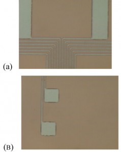
Figure 2: Photomicrographs of ITO: (a) bonding pads and leads (2µm wide) and (b) sensing electrode pads (20 µm x 20 µm).
Accordingly, the Fonstad and Boyden groups, in a collaborative effort, have set out to devise and test strategies for (1) coating metal electrodes to make them insensitive to light when immersed in the brain and (2) using standard photolithographic and micro-fabrication techniques to produce linear arrays of light-proof electrodes for high-density recording of neural spikes and field potentials (in the style of the “Michigan probe”). For both of these strategies, the transparent conductor indium tin oxide (ITO) is being used. Dense linear arrays of electrodes have been fabricated from ITO-coated substrates. The electrodes have impedances (at 1 kHz) of 1 megaohm, with no detectable light artifact. Techniques are now being developed to fashion linear insertable probes with many (e.g., 40) light-proof ITO recording sites while maintaining a thin, tissue-damage-minimizing (e.g., 150 × 50 micron) cross-section. Furthermore, the electrode substrate is polyethylene terephthalate (PET), a transparent polymer often used in the flexible OLED community. This choise in substrate satisfies the need for optical insensitivity while reducing the modulus gradient between implant and neural cells. As high modulus implants tend to induce cell death as well as become engulfed in scar material, this characteristic is important and beneficial.
Such novel probes will allow artifact-free recordings during optical neuro-modulation, enabling the systematic analysis of real-time neural dynamics across multiple time scales, via causal neural control tools. They may also be important for assessing the effects of optical neural control on the treatment of intractable brain disorders.
References
- X. Han and E.S. Boyden, “Multiple-Color Optical Activation, Silencing, and Desynchronization of Neural Activity, with Single-Spike Temporal Resolution,” PLoS ONE vol. 2, no,. 3, p. e299, March 2007. [↩]