Flexible multi-site electrodes for moth flight control

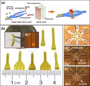
Figure 1: (a) Schematic of the probe, showing the split-ring design that enables the probe to encircle the nerve cord. (b) Image showing various FME designs. Close-up images on the split-ring structure of the (c) WH6, (d) BC6, and (e) BC8 designs.
Significant interest exists in creating insect-based Micro-Air-Vehicles (MAVs) [1] [2] [3] that would combine advantageous features of insects—small size, effective energy storage, navigation ability—with the benefits of MEMS and electronics—sensing, actuation and information processing. The key part of the insect-based MAVs is the stimulation system which interfaces with the nervous system of the insect to bias the insect’s flight path.

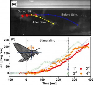
Figure 2: (a) Side view image of a freely flying moth that has been stimulated to perform right turns following the elicited abdominal motions (repeatable for 4 successive stimulations). The locations of the moth before, during and after the stimulation are marked by the triangle, circle and square, respectively. (b) The changes of the yaw angle (θ) of the moth with 4 successive stimulations.
In this work, we have developed a flexible multi-site electrode (FME) for insect flight control that directly interfaces with the animal’s central nervous system. The FMEs are made of two layers of polyimide with gold sandwiched in-between in a split-ring geometry using standard MEMS processing [3]. The FMEs have a novel split-ring design that incorporates the anatomical bi-cylinder structure of the nerve cord of the Moth Manduca Sexta and allows for an efficient surgical process for implantation (Figure 1). Additionally, we have integrated carbon nanotube (CNT)-Au nanocomposites into the FMEs to enhance the charge injection capability of the electrode.
We are able to elicit graded and multi-direction abdominal movements in both the pupae and adult moths using FME stimulation.
Moreover, the CNT coated FMEs are able to elicit abdominal motion of the moths with a stimulation voltage significantly less (1.0 V vs. 2.0 V, p < 0.001, n=10 moths) than that of uncoated FMEs. Finally, we have integrated the FMEs into a wireless system and in the flight control experiment, we are able to force a freely flying animal to perform turning motions (Figure 2a) using the abdominal ruddering with these elicited abdomen motions. These turning motions are well repeatable and the changes in the yaw angle of the moth with 4 successive stimulations are shown in Figure 2b.
References
- A. Bozkurt, F. Gilmour, A. Lal, “Balloon assisted flight of radio-controlled insect biobots,” IEEE Transactions on Biomedical Engineering, Vol 56, pp. 2304-2307, Sept. 2009. [↩]
- H. Sato, C. W, Berry, Y. Peeri, E. Baghoomian, B. E. Casey, G. Lavella, J. M. VandenBrooks, J. F. Harrison and M. M. Maharbiz, “Remote radio control of insect flight,” Frontiers in Integrative Neuroscience, vol 3, pp. 1-11, Oct 2009. [↩]
- W.M. Tsang, A. Stone, Z. Aldworth, D. Otten, T. Akinwande, T. Daniel, J.G. Hildebrand, R. Levine, J. Voldman, “Remote Control of A Cyborg Moth Using Carbon Nanotube-Enhanced Flexible Neuroprosthetic Probe,” in Proc. IEEE MEMS2010, pp. 39-42, Jan. 2010. [↩] [↩]