Cell-based Sensors for Measuring Impact of Microsystems on Cell Physiology
- Category: MEMS & BioMEMS
- Tags: catherine lo, joel voldman
The use of microsystems to manipulate and study cells in microenvironments is continually increasing. However, along with such increase in usage comes a growing concern regarding the impact of these microsystems on cell physiology. In this project, we are developing a set of cell-based fluorescent sensors to measure the impact of common stresses experienced in microsystems on cell physiology. We are including stress agents commonly found in microsystems (e.g., UV exposure, heat shock, fluid flow, etc.). Each sensor is designed to respond to one particular stress agent but can also be combined for multiplexed analysis of multiple stresses at once, as might be experienced in a typical microsystem. Each sensor will use different colors to both indicate the type of sensor and the strength of the signal, to ease multiplexed analysis.
We are currently developing a sensor that responds to activation of the p53 protein pathway, for generalized DNA damage analysis. Similar to the heat shock sensor we previously reported [1] [2] , which coupled fluorescent protein expression to activation of heat shock factor 1, the DNA damage sensor will couple fluorescent protein expression to activation of p53. The new sensor will have cyan (cerulean) as the constitutive color and red (RFP) as the activation color. Figure 1 shows the DNA damage sensor response after 30 min of UV exposure. One of the more relevant sources for physiological stress on cells cultured in microfluidic devices is shear stress. Construction of a shear stress sensor cell line requires an understanding and characterization of the gene expression mechanisms and mechanotransduction pathways, especially since the pathways are known to have a varying correlation towards cell types, magnitudes, and dynamics of applied stresses. Therefore, we are using a multi-flow microfluidic device (see Figure 2) that can simultaneously apply different flows to cells across a 1000× range to first understand the behavior of NIH3T3 mouse fibroblast cells under flow [3] . Specifically, these cells are seeded in 6 chambers concurrently, exposed to flow for 1-6 hours, and assayed for gene expression changes. Once they are characterized, we will construct a transfected NIH3T3 cell line with RFP expression correlating to shear stress.
-
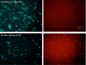
- Figure 1: DNA Damage Sensor. Images of NIH3T3 cells (A) before and (B) after UV exposure for 30 min and recovery for 4 hrs. The left images are of the constitutive cyan reporter while the right images are of the red fluorescent DNA damage reporter.
-
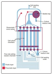
- Figure 2: Multi-flow microfluidic device. Shown is a schematic of the multi-flowrate microfluidic device, showing the fluidic layer (blue) where the cells are grown and the pneumatic layer (red) containing microvalves.

- S. P. Desai and J. Voldman, “Cell-based sensors for quantifying the physiological impact of microsystems,” Integrative Biology, vol. 3, pp. 48-56, 2011. [↩]
- S. P. Desai and J. Voldman, “Measuring the impact of dielectrophoresis on cell physiology using a high-content screening platform,” in Micro Total Analysis Systems, 2008, pp. 1308-10. [↩]
- Y.-C. Toh and J. Voldman, “Fluid shear stress primes mouse embryonic stem cells for differentiation in a self-renewing environment via heparan sulfate proteoglycans transduction,” Faseb Journal, vol. 25, pp. 1208-17, 2011. [↩]