High Channel-count Silicon Neural Recording Probes for 3-D Characterization of Neural Dynamics
- Category: Medical Electronics, Optics & Photonics
- Tags: Clifton Fonstad
Optogenetics is commonly used for precision modulation of the activity of specific neurons within neural circuits [1] , but assessing the impact of optogenetic neural modulation on millisecond-timescale local and global circuit neural activity remains difficult. We have developed a novel strategy for designing and fabricating silicon-based microelectrode arrays with customizable electrode locations, targetable to defined neural substrates distributed in a 3-D pattern throughout a neural network in the mammalian brain, and compatible with simultaneous use of a diversity of existing light delivery devices. Our design of these 3-D electrode arrays provides for both easy electrical and mechanical assembly, and provides for scaling of arrays to up to 1000 neural recording channels and beyond.
Our approach relies upon a number of innovations at the material, structural, electrical, and data acquisition levels. First, typical silicon-based electrodes that are arranged in a 1-dimensional linear array, or 2-dimensional comb-like fashion, often use linear or tetrode-style electrode locations along the comb’s fingers, with stereotyped spacing and pad sizes. Our software-driven approach enables variable spacing and pad sizes, so that electrode geometries can be customized to the cellular properties of the brain circuits under investigation. Second, to support the assembly of such electrode arrays into a 3-dimensional array, we have developed novel electrical and mechanical connector strategies to make the assembly as automated and reliable as possible. Third, we have developed strategies for amplifying and acquiring data that simplify the use of these probes in an intact, in vivo, mammalian context. Fourth, we have implemented hybrid electrodes that contain both a low-impedance metallic pad for recording of spike activity, as well as an indium tin oxide (ITO) pad that can report local field potentials (LFPs) without the photo-electrochemical artifacts common in optogenetics. Finally, these 3-D probes are designed to be easy to use, from design to surgery. We have developed a user-friendly interface that enables neuroscientists to specify probe geometries based upon neural target geometries and coordinates, and are developing supporting surgical and behavioral strategies for use of such arrays in vivo.
-
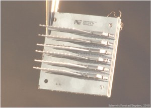
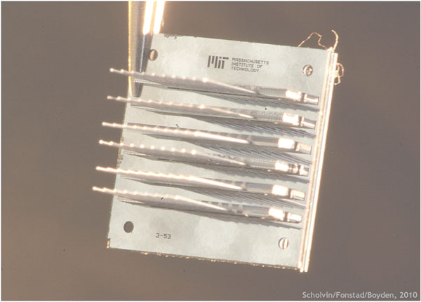
- Figure 1: A photomicrograph of six ten-probe linear arrays assembled into a six by ten two-dimensional array of probes. The probes in this figure and in Figure 2 were fabricated for assembly studies and do not have sense electrodes.
-
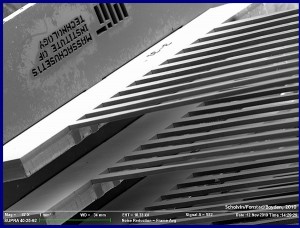
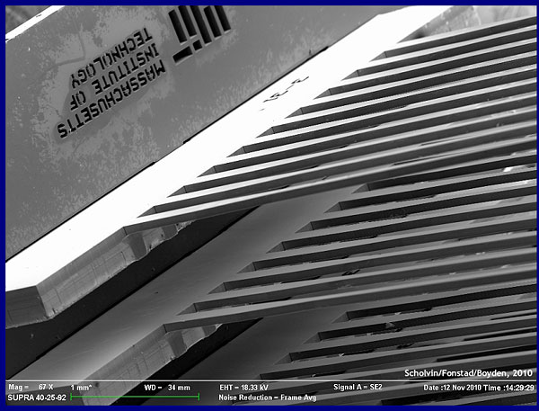
- Figure 2: A close-up scanning electron micrograph of a portion of an array like that in Figure 1. The probes in the linear array are spaced 400 µm center-to-center, and the linear arrays are spaced 800 µm
- X. Han and E. S. Boyden, “Multiple-color optical activation, silencing, and desynchronization of neural activity, with single-spike temporal resolution,” PLoS ONE, vol. 2, no. 3, p. e299, Mar. 2007. [↩]